Echocardiography
Echocardiography
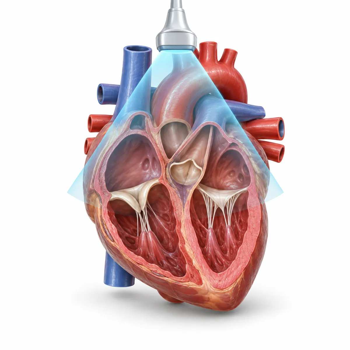
What it is
A non-invasive ultrasound examination that visualises the heart chambers, valves, muscle walls and blood flow in real time. Valve function, myocardial contractility, chamber dimensions and ejection fraction are assessed. No X-ray radiation is used and no special preparation is required.
Why it is performed
Echocardiography evaluates the function of the heart valves (stenosis, insufficiency), the contractile ability of the myocardium, chamber sizes, and ejection fraction (the percentage of blood the heart pumps out with each contraction). These indicators play a key role in detecting heart failure, valve diseases, cardiomyopathies, and other structural changes.
How it is performed
The patient lies comfortably on an examination table, a special gel is applied to the chest, and an ultrasound probe is moved across the chest to capture images of the heart from various angles. The examination usually takes 20-30 minutes and causes no discomfort to the patient.
Who may be recommended this examination
A physician may recommend this examination to patients with heart murmurs, shortness of breath, chest pain, changes in heart rhythm, arterial hypertension, suspected heart failure, or those requiring regular cardiac monitoring.
Preparation
Echocardiography usually requires no special preparation — there is no need to fast or follow a diet beforehand. Comfortable clothing is sufficient.
Results
The results of the examination are typically interpreted by a cardiologist immediately after the procedure, and the findings are used to determine further treatment strategy.